- Products & Services TokuProfile Spectral Flow CytometryTokuKit
- Resources
- Pricing
- Company
- Login
FAQ
What is a state marker?
State markers are specific proteins that indicate the functional state or condition of a cell at a point in time. Some examples of different states of cells include activation, exhaustion, and proliferation. Some state markers on our standard Peripheral Blood Mononuclear Cell (PBMC) panel are Ki67, a protein all cells produce when replicating, and PD-1, a checkpoint protein expressed on exhausted T cells.
For each cell analyzed, we measure the level of expression of all 25+ proteins on our panel, including all state markers. Data for state markers included in our panels and our web app are reported as frequency and a median channel value (MCV). The frequency represents the proportion of cells within each cell subset expressing that state marker above a set threshold. We say these cells are “positive” for this state marker. This can answer the question “What proportion of T cells are exhausted?” with “37% of T cells are positive for PD-1 and therefore are exhausted.”
The MCV represents the intensity of the signal detected for a specific metal isotope associated with a particular antibody tag on a cell. It is similar to median fluorescence intensity, or MFI, in flow cytometry. This can answer the question “Does the amount of PD-1 expressed on T cells increase or decrease with treatment?” with “The amount of PD-1 expressed on T cells decreases with treatment because the MCV value for PD-1 on T cells drops from 5.6 to 4.8 after treatment.” This type of marker expression level information is important since higher PD-1 protein expression correlates with a more exhausted phenotype (PNAS 2013).
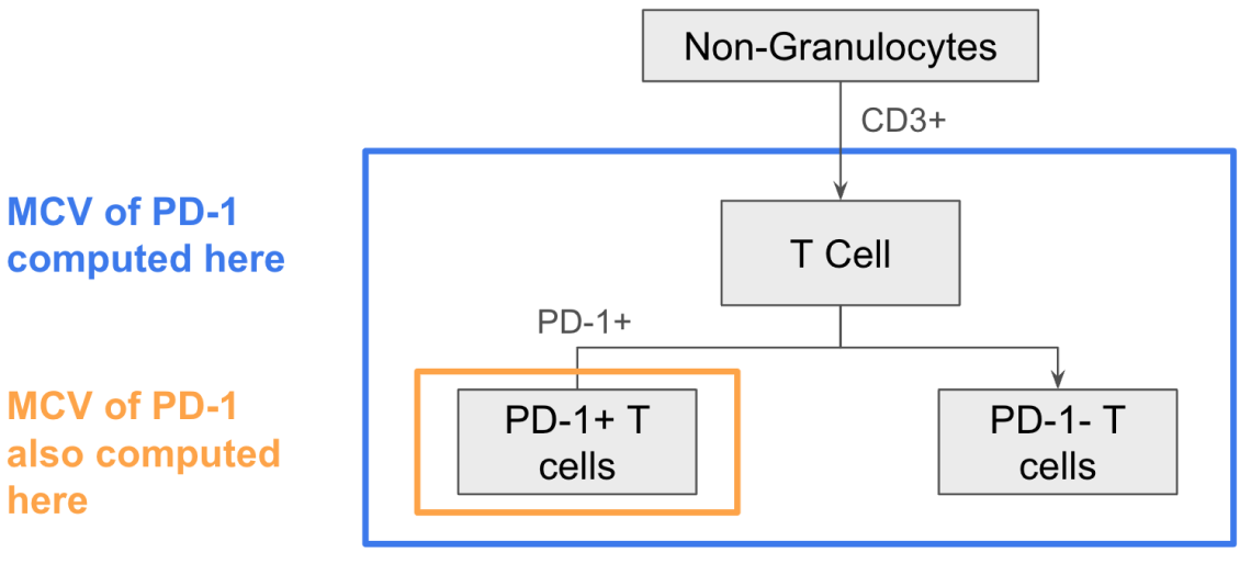
We evaluate state marker expression as an MCV for a given marker in the (1) marker-positive cell population and (2) parent population. This allows you to quantify the level of expression within cells that are known to express the state marker of interest and across the entire parent population, including cells with no or low expression of the state marker.